- Advanced imaging technologies can reveal the intricate ecosystem surrounding glioblastoma tumors, offering new insights into treatment resistance.
- Glioblastoma’s highly adaptive tumor microenvironment actively suppresses immune responses and fosters treatment resistance.
- Glioblastomas extend tendrils deep into brain tissue, making complete surgical removal nearly impossible.
- Immunosuppressive cells like regulatory T cells and tumor-associated macrophages protect cancer cells from immune attack.
- Hypoxia within the tumor core drives genetic instability and treatment resistance in glioblastoma.
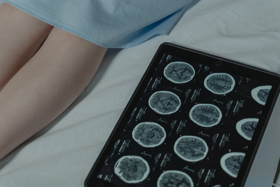
Why do glioblastomas resist nearly every available treatment, leading to grim survival rates despite decades of research? This question haunts neuro-oncologists and patients alike. Glioblastoma, the most aggressive form of primary brain cancer, claims most lives within 15 months of diagnosis. Traditional therapies—surgery, radiation, and chemotherapy—often fail because the tumor infiltrates healthy brain tissue and evolves rapidly. But emerging evidence suggests that part of the answer lies not just in the tumor cells themselves, but in the intricate ecosystem surrounding them: the tumor microenvironment. Now, advanced imaging technologies are offering an unprecedented window into this hidden world, revealing how immune cells, blood vessels, and metabolic activity interact to shield the tumor from destruction.
\n\n
What Makes Glioblastoma So Resistant to Treatment?

\n
The resilience of glioblastoma stems from its highly adaptive tumor microenvironment, which actively suppresses immune responses and fosters treatment resistance. Unlike many cancers that form discrete masses, glioblastomas extend tendrils deep into brain tissue, making complete surgical removal nearly impossible. Moreover, the microenvironment surrounding the tumor is rich in immunosuppressive cells like regulatory T cells and tumor-associated macrophages, which protect cancer cells from immune attack. Hypoxia—low oxygen levels within the tumor core—further drives genetic instability and resistance to radiation. Recent studies using multiparametric MRI and positron emission tomography (PET) have revealed that metabolic reprogramming, such as increased glucose uptake via the Warburg effect, allows tumor cells to thrive under stress. These insights, made visible through advanced imaging, are reshaping how clinicians understand and approach treatment.
\n\n
What Evidence Supports the Role of Imaging in Glioblastoma Care?

\n
Clinical studies using techniques like amide proton transfer (APT) MRI and amino acid PET scans have demonstrated their ability to map tumor metabolism and cellular density in real time. A 2023 study published in Nature Medicine showed that APT MRI could distinguish between treatment-related inflammation and true tumor recurrence with over 85% accuracy—critical for avoiding unnecessary surgeries. Meanwhile, PET imaging with tracers like FET (fluoroethyltyrosine) allows clinicians to visualize active tumor regions beyond what standard MRI detects. At the University of California, San Francisco, researchers combined these modalities with single-cell sequencing to identify spatial patterns of immune evasion. According to the CDC, glioblastoma accounts for nearly half of all malignant brain tumors, underscoring the urgency of integrating imaging advances into routine care. These tools are not just diagnostic—they’re guiding targeted therapies and clinical trial design.
\n\n
Are There Limitations to These Imaging Advances?

\n
Despite their promise, advanced imaging techniques face challenges in accessibility, cost, and interpretation. Many hospitals lack the specialized equipment or trained personnel needed to perform and analyze APT MRI or amino acid PET scans. Furthermore, the data generated are highly complex, requiring computational models and expert radiologists to extract meaningful insights. Some experts caution that overreliance on imaging biomarkers could lead to premature treatment changes without sufficient clinical validation. Additionally, tumor heterogeneity means that even high-resolution images may miss microscopic pockets of resistance. There’s also ethical concern about patient burden—repeated scans expose individuals to radiation and prolonged MRI sessions, which can be taxing for those with neurological symptoms. While these technologies represent a leap forward, they’re not yet a standalone solution and must be integrated carefully within multidisciplinary care frameworks.
\n\n
How Are Patients Benefiting from These Innovations Today?

\n
Patients are already seeing tangible benefits through more accurate diagnoses and personalized treatment plans. At leading cancer centers like MD Anderson and Mayo Clinic, imaging-guided biopsies ensure that tissue samples are taken from the most aggressive tumor regions, improving diagnostic precision. In clinical trials, real-time imaging is being used to assess response to immunotherapies and targeted drugs, allowing for early adjustments. One patient in a 2022 trial at Johns Hopkins received a customized vaccine based on imaging and genomic data, resulting in a 70% reduction in tumor volume over six months. Beyond treatment, advanced imaging helps families understand disease progression more clearly, supporting informed decision-making. As these tools become more standardized, they could reduce diagnostic delays and improve survival outcomes across broader populations.
\n\n
What This Means For You
\n
If you or a loved one is facing a glioblastoma diagnosis, emerging imaging technologies offer hope for more precise and effective care. These tools can help distinguish between tumor growth and treatment effects, reduce unnecessary interventions, and guide access to cutting-edge therapies. While not universally available yet, their integration into major medical centers is accelerating. Staying informed and seeking care at institutions with advanced neuroimaging capabilities may significantly impact treatment outcomes.
\n\n
As imaging continues to evolve, a critical question remains: Can we use these dynamic maps of the tumor microenvironment to predict treatment response before therapy even begins? Answering this could shift glioblastoma care from reactive to proactive, transforming a once uniformly fatal diagnosis into a manageable condition.
Source: MedicalXpress