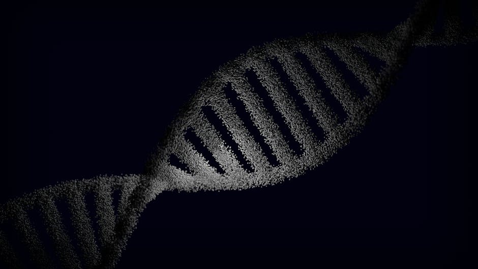
- Three-dimensional DNA folding in developing B cells plays a crucial role in preventing autoimmune diseases by inactivating self-reactive genetic sequences.
- Approximately 98% of B cells with self-reactive potential are silenced through structural DNA changes, rather than cell death alone.
- Research suggests that disrupting the DNA folding mechanism may underlie autoimmune conditions such as lupus and type 1 diabetes.
- Genome topology, specifically the folding of DNA, may hold the key to new diagnostic and therapeutic strategies for autoimmune diseases.
- In mouse models, researchers found that aberrant DNA folding increased self-reactive antibody expression by 8.5-fold in the absence of the architectural protein CTCF.
Recent research reveals that precise three-dimensional DNA folding in developing B cells plays a pivotal role in preventing the immune system from producing antibodies that attack the body’s own tissues. This mechanism, which operates during antibody gene rearrangement, selectively inactivates self-reactive genetic sequences by altering chromatin architecture. Disruption of this process may underlie autoimmune conditions such as lupus and type 1 diabetes, suggesting new diagnostic and therapeutic strategies rooted in genome topology.
\n\n
DNA Rearrangement and Immune Tolerance: The Data

\n
Published in Nature, the study demonstrates that during V(D)J recombination—a process where gene segments are shuffled to generate diverse antibodies—nearly 98% of B cells with self-reactive potential are silenced through structural DNA changes rather than cell death alone. Using high-resolution chromatin conformation capture (Hi-C) and single-cell RNA sequencing in mouse models, researchers mapped how specific genomic regions fold into insulated neighborhoods, physically separating self-reactive variable (V) gene segments from recombination machinery. In mutant mice lacking the architectural protein CTCF, aberrant folding increased self-reactive antibody expression by 8.5-fold. These findings were corroborated in human B cell precursors, where similar topological boundaries were disrupted in samples from patients with systemic lupus erythematosus, suggesting conserved mechanisms across species.
\n\n
Key Players: B Cells, CTCF, and the V(D)J Machinery

\n
The primary actors in this immune safeguard are developing B lymphocytes, which undergo rigorous selection in the bone marrow to eliminate self-reactive clones. Central to this process is the CCCTC-binding factor (CTCF), a protein known for its role in shaping chromatin loops. In normal B cell development, CTCF binds to specific insulator sequences flanking antibody variable gene clusters, forming loop domains that restrict access to certain V genes. When these boundaries are compromised, normally sequestered self-reactive V segments become available for recombination. The RAG endonuclease complex, responsible for cutting and splicing DNA during V(D)J recombination, gains access to these regions, inadvertently enabling the creation of autoantibody-encoding genes. Researchers also identified reduced expression of cohesin subunits in autoimmune-prone individuals, further implicating structural genome regulators in immune tolerance.
\n\n
Trade-Offs: Diversity Versus Self-Tolerance

\n
The immune system faces a fundamental trade-off: maximizing antibody diversity to combat countless pathogens while minimizing the risk of self-reactivity. This study highlights how 3D genome organization resolves this tension—not by eliminating all self-reactive sequences, which would drastically reduce repertoire breadth, but by spatially controlling their accessibility. The benefit is a highly diverse yet largely self-tolerant antibody pool. However, this system carries inherent risks; mutations in CTCF binding sites or dysregulation of chromatin remodelers can erode these protections, leading to autoimmune disease. Conversely, overzealous silencing might limit responses to certain infections. Therapeutically, modulating chromatin architecture could enhance vaccine responses or correct autoimmune defects, but such interventions must avoid destabilizing the delicate balance between immunity and tolerance.
\n\n
Timing: Why This Discovery Emerges Now

\n
This breakthrough follows a decade of advances in spatial genomics and single-cell technologies that have made it possible to observe dynamic chromatin folding in rare cell populations like early B cells. Previous models assumed that self-reactive B cells were primarily removed through apoptosis or receptor editing. However, the inability to fully explain the scale of immune tolerance prompted researchers to explore epigenetic and structural mechanisms. The convergence of CRISPR-based genome engineering, improved Hi-C protocols, and autoimmune disease genomics allowed the team to test the functional impact of specific folding disruptions. The identification of non-coding mutations in CTCF sites among lupus patients provided the clinical correlation that solidified the findings’ relevance.
\n\n
Where We Go From Here
\n
In the next 6 to 12 months, three scenarios could unfold: First, pharmaceutical companies may begin screening for small molecules that stabilize CTCF-mediated loops, aiming to treat early-stage autoimmunity. Second, clinical labs might incorporate chromatin topology markers into diagnostic panels for high-risk patients, enabling earlier intervention. Third, gene therapy approaches could be developed to correct pathogenic folding defects in hematopoietic stem cells, particularly for monogenic autoimmune disorders. Each path depends on confirming these mechanisms in broader human cohorts and ensuring that manipulating genome structure does not inadvertently promote malignancies such as B cell lymphoma, where V(D)J errors are already implicated.
\n\n
Bottom line — the discovery that DNA folding acts as a checkpoint in antibody gene assembly transforms our understanding of immune tolerance, positioning genome architecture as a central player in preventing autoimmune disease.
Source: Nature