- Researchers have created a comprehensive set of reference charts for white matter development and aging across the human lifespan.
- This dataset of 35,120 brain scans, sourced from 124 studies across six continents, provides unprecedented insights into white matter changes.
- The brain charts can be used as a benchmark to track individual deviations, potentially signaling early neurological disease.
- The lack of universal standards for brain development has hindered clinical diagnosis and research into neurodevelopmental and neurodegenerative disorders.
- White matter changes influence cognition, motor control, and emotional regulation throughout the lifespan.
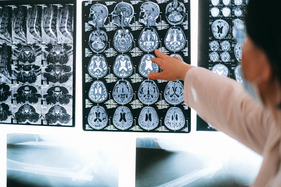
For the first time in neuroscience, researchers have constructed a complete set of reference charts mapping the development and aging of white matter in the human brain across the entire lifespan—based on an unprecedented dataset of 35,120 brain scans. Published in Nature on May 13, 2026, this global initiative integrates neuroimaging data from 124 studies across six continents, enabling scientists and clinicians to track normative changes in both the microscopic (microstructural) and large-scale (macrostructural) properties of white matter. These brain charts function much like pediatric growth curves, offering a benchmark against which individual deviations—potentially signaling early neurological disease—can now be compared with remarkable precision.
\n
The Urgent Need for Standardized Brain Metrics

\n
Until now, the absence of universal standards for brain development has hindered both clinical diagnosis and research into neurodevelopmental and neurodegenerative disorders. While pediatricians have relied on growth charts for height, weight, and head circumference for decades, equivalent tools for brain maturation have been fragmented or limited to narrow age ranges. White matter, the neural wiring that connects brain regions, undergoes dynamic changes from fetal development through aging, influencing cognition, motor control, and emotional regulation. Disruptions in its development are linked to conditions such as autism, multiple sclerosis, and Alzheimer’s disease. The new study addresses a critical gap by synthesizing decades of disparate MRI data into unified, age-stratified trajectories, enabling earlier detection of abnormalities and more accurate tracking of disease progression.
\n
Global Collaboration Powers Unprecedented Dataset

\n
The landmark study was led by an international consortium of neuroscientists, radiologists, and data scientists from institutions including the University of Edinburgh, Harvard Medical School, and the Max Planck Institute for Human Cognitive and Brain Sciences. By pooling anonymized diffusion MRI and structural MRI data from 35,120 individuals aged 0 to 100, the team constructed percentile curves for key white matter properties: fractional anisotropy (a measure of microstructural integrity), mean diffusivity (reflecting tissue density), and total white matter volume. Data were harmonized using advanced computational algorithms to account for differences in scanner types, protocols, and populations, ensuring robustness across diverse cohorts. The resulting charts reveal distinct phases of development—rapid myelination in early childhood, peak integrity in early adulthood, and gradual decline after age 50.
\n
Revealing the Lifespan Trajectory of Brain Connectivity

\n
The analysis uncovered non-linear trajectories in white matter development, with the most dramatic changes occurring in the first two years of life. Microstructural complexity increases sharply during infancy, coinciding with language acquisition and motor skill development. Macrostructural volume peaks around age 30, after which it declines at an accelerating rate post-60. Notably, the study found that individual variability in white matter structure increases with age, suggesting divergent aging pathways. Certain brain tracts, such as the corpus callosum and superior longitudinal fasciculus, showed earlier maturation and later degeneration, aligning with known cognitive aging patterns. These findings offer a granular view of how brain connectivity evolves, providing a foundation for identifying atypical patterns before symptoms manifest.
\n
Implications for Early Diagnosis and Precision Medicine

\n
The clinical implications of these brain charts are profound. Pediatric neurologists can now compare a child’s white matter development to population norms, potentially flagging developmental delays years before traditional assessments. In aging populations, deviations from expected decline could signal early neurodegeneration, enabling earlier intervention in diseases like Alzheimer’s. The charts may also improve clinical trial design by stratifying participants based on brain age rather than chronological age, increasing sensitivity to treatment effects. Furthermore, the open-access nature of the dataset and modeling tools allows global researchers to refine the charts and extend them to specific populations, such as those with genetic risk factors or preterm birth histories.
\n
Expert Perspectives
\n
“This is a milestone in quantitative neuroscience,” said Dr. Sarah Whitaker, a neuroimaging expert at University College London not involved in the study. “We finally have a reference framework akin to the Framingham Heart Study, but for the brain.” However, some researchers urge caution. Dr. Kenji Tanaka of Kyoto University warns that “population averages may obscure important ethnic, socioeconomic, or environmental influences on brain development.” While the dataset is globally diverse, underrepresentation from low-income regions remains a limitation, potentially affecting the universality of the charts.
\n
Looking ahead, the consortium plans to incorporate functional MRI and genetic data to explore the biological drivers of white matter trajectories. Future updates may include sex-specific charts and dynamic models that account for lifestyle factors like exercise, sleep, and education. As artificial intelligence tools evolve, these brain charts could be integrated into routine clinical imaging software, providing real-time assessment of brain health. The next frontier lies in translating these population-level insights into personalized neurological care—ushering in an era where brain development is not just observed, but actively monitored and optimized.
Source: Nature